
Home / News / Industry News / Common Causes of Upper Airway Obstruction and How Clinicians Spot Them
Content
A patient becomes restless during sedation, then quiet, then difficult to ventilate. The first question is not “which rare disease is this?” but “what is blocking airflow right now?” In most acute settings, the common causes of upper airway obstruction are practical and often reversible: the tongue falling back when consciousness drops, secretions or vomit pooling in the pharynx, soft tissue swelling, trauma, a foreign body, or laryngospasm. The cause may be simple, but the consequence is not; obstruction can progress from noisy breathing to silent hypoxemia within minutes.
The clinical setting usually narrows the list faster than a long differential. An unconscious patient in the emergency department points first to tongue displacement and secretions. A child with fever and drooling points to infection. A patient who chokes while eating points to a foreign body. A postoperative patient with neck swelling points to hematoma or edema. A slow, progressive history with hoarseness or exercise limitation points to structural narrowing or a mass. This article groups the common causes by mechanism, then shows how setting, onset, and bedside clues change the working diagnosis.
Upper airway obstruction is better understood by mechanism than by a flat list. The same symptom—stridor, snoring, or increased work of breathing—can come from a floppy tongue, a swollen epiglottis, a narrowed trachea, or a functional problem such as laryngospasm. Grouping causes this way supports a faster airway plan and a more sensible device choice.
| Mechanism | Common causes | Typical clues |
|---|---|---|
| Loss of muscle tone | Tongue fallback, sedation, opioid effect, stroke, seizures, cardiac arrest | Snoring, poor air entry, improvement with jaw thrust or repositioning |
| Fluid or material in the pharynx | Vomitus, blood, secretions, mucus plugs | Gurgling, visible pooling, suction improves ventilation |
| Soft tissue swelling or infection | Epiglottitis, supraglottitis, croup, peritonsillar or retropharyngeal abscess, angioedema, post-extubation edema | Fever, drooling, muffled voice, rapid onset, positional preference, stridor |
| Mechanical blockage | Food, bones, teeth, dentures, toys, foreign body in nose or pharynx | Sudden onset while eating or playing, coughing, choking, unilateral noise |
| Trauma or compression | Laryngeal injury, facial fractures, neck hematoma, burns, inhalation injury, goiter or mass | History of trauma or surgery, neck swelling, hoarseness, bruising, progressive stridor |
| Functional or structural narrowing | Laryngospasm, vocal cord paralysis, subglottic stenosis, tracheal stenosis, tumors, obesity-related collapse | Voice change, exertional symptoms, previous intubation or neck surgery, sleep-related obstruction |
Onset speed is one of the most useful discriminators. Sudden obstruction in an otherwise alert adult should raise suspicion for a foreign body, laryngospasm, angioedema, or vomit. Rapid progression over hours suggests infection, edema, or hematoma. A slower course over weeks suggests a mass, goiter, stenosis, or vocal cord problem. A patient who is already unconscious cannot report symptoms, so the examination must focus on air movement, chest rise, voice, swallowing, and the effect of simple repositioning.
Age also shifts the probability. In children, croup, epiglottitis, foreign bodies, laryngomalacia, and congenital narrowing are more prominent. In adults, obstructive sleep apnea, obesity, tumors, vocal cord paralysis after thyroid or neck surgery, and post-extubation edema are more common. In older adults, dentures, stroke, dementia, and sedating medicines add aspiration and tongue-fallback risk. A cause that is rare in one group may be the first thing to exclude in another.
The presence of fever, drooling, and difficulty swallowing matters. A child who sits forward and refuses to lie flat should not be forced supine for a routine examination; the position may be protecting a swollen airway. In contrast, a patient who improves immediately with a jaw thrust or head tilt has a functional component, not a fixed structural blockage. These distinctions change urgency, equipment, and the safest approach to securing the airway.
In infants and young children, the airway is smaller and more compliant, so a modest amount of swelling can cause disproportionate obstruction. Croup produces a barking cough and inspiratory stridor. Epiglottitis and supraglottitis can cause fever, drooling, and a muffled voice. Foreign bodies are often food, nuts, or small toys, and the history may be missing because the event was not witnessed. Congenital causes such as laryngomalacia, tracheal stenosis, and vascular rings may present with feeding difficulty, recurrent noisy breathing, or poor weight gain.
Adults more often present with acquired problems. Obesity and obstructive sleep apnea cause repeated pharyngeal collapse during sleep. Laryngeal or thyroid tumors can narrow the airway gradually while producing hoarseness or a change in voice. Vocal cord paralysis may follow thyroid, carotid, or cervical spine surgery. Angioedema from medications or allergic reactions can develop quickly and involve the tongue, lips, and supraglottic tissues. Trauma from facial fractures, laryngeal injury, or inhalation burns can worsen as edema develops.
In anesthesia and critical care, some of the most common causes of upper airway obstruction are created or worsened by the clinical intervention itself. Sedatives and opioids reduce muscle tone and blunt protective reflexes. Supine positioning encourages the tongue and soft palate to fall backward. Blood, saliva, and gastric contents can accumulate above the glottis. Laryngospasm can close the vocal cords reflexively after light anesthesia, irritation, or secretions. Post-extubation edema, hematoma, and tube-related injury can narrow the airway after a procedure that initially appeared uneventful.
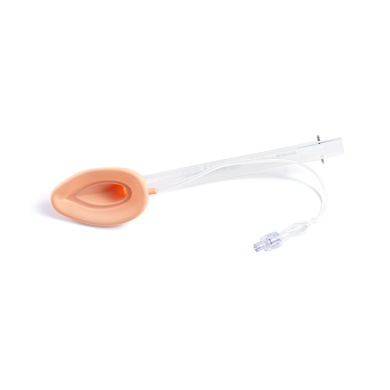
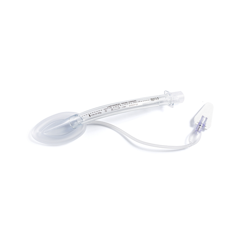
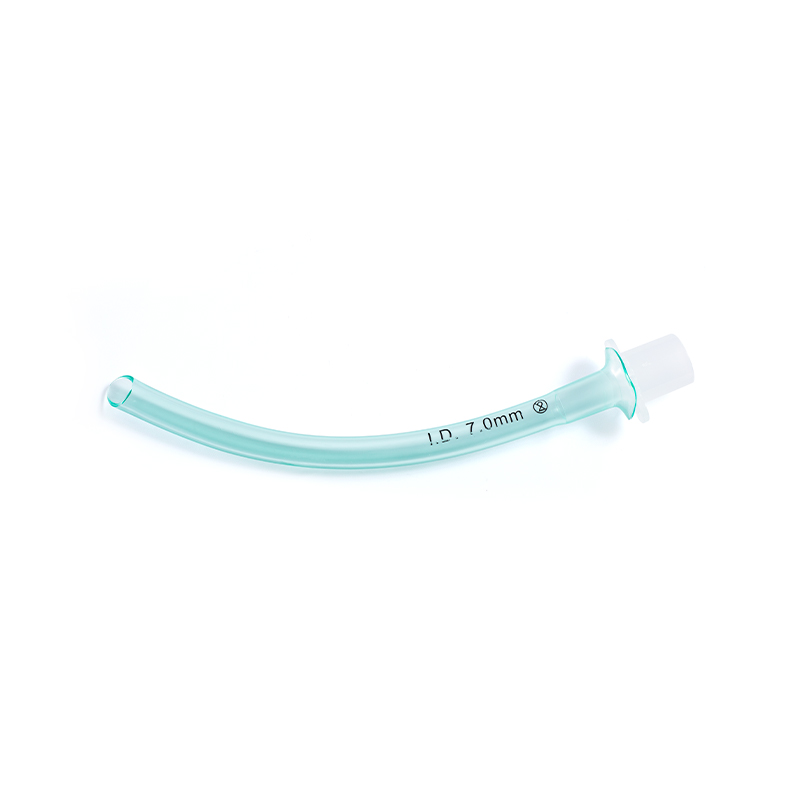
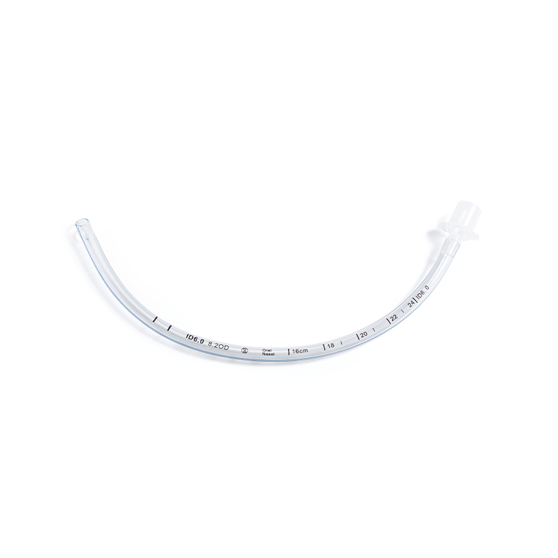
Device selection should follow the cause and the planned duration of support. A patient with poor consciousness but preserved reflexes may need a nasopharyngeal airway or careful positioning before more advanced support. A patient with a foreign body needs removal, not simply a bypass. A patient with supraglottic swelling may need a smaller-than-usual tube, a bougie, or surgical backup. In rescue ventilation, a laryngeal mask airway can be useful when face-mask ventilation is difficult, especially when a gastric access channel is needed to reduce aspiration risk. For planned intubation, a cuffed tube with careful placement and ongoing cuff pressure monitoring helps limit pressure-related injury.
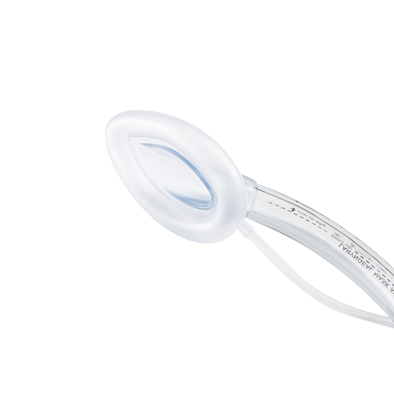 Standard PVC Laryngeal Mask AirwayDisposable supraglottic airway with sterile single-use packaging and sizes from neonate to adult, useful when rescue ventilation is needed and face-mask ventilation is difficult.View Product →
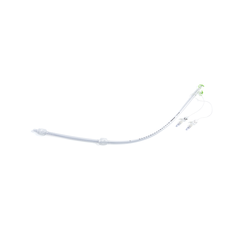
Product design alone does not solve an airway emergency, but familiar, well-matched equipment reduces delay. A standard PVC laryngeal mask airway, a positioning-friendly cuffed endotracheal tube, and a cuff pressure monitoring system each address a different part of the same problem: maintaining a patent airway while limiting avoidable trauma. Clinicians should confirm sizing, check the cuff, prepare suction, and have a plan for failed ventilation before the first attempt.
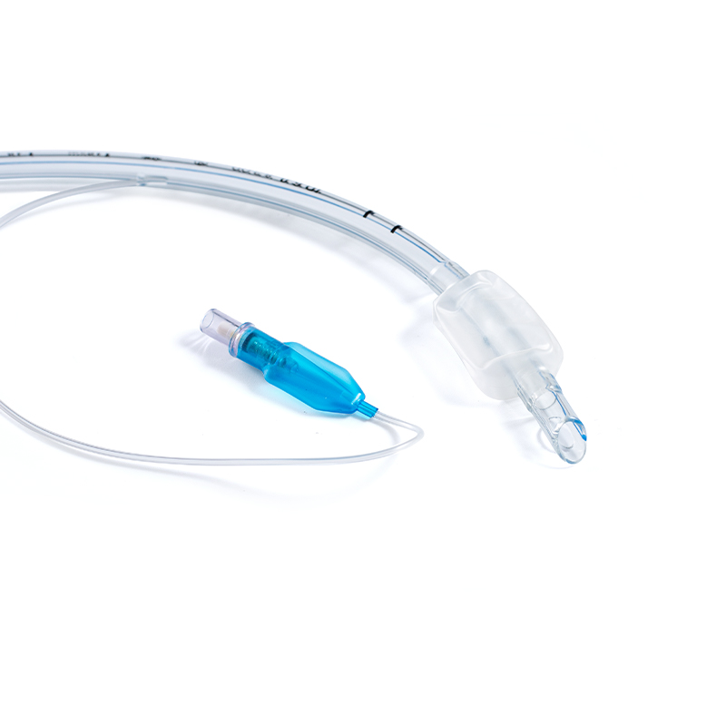 Standard Cuffed Endotracheal Tube with Depth MarkingsSingle-use cuffed tube with clear depth scales, a low-pressure high-volume cuff, and adult and pediatric sizes for planned intubation and ventilation support.View Product →
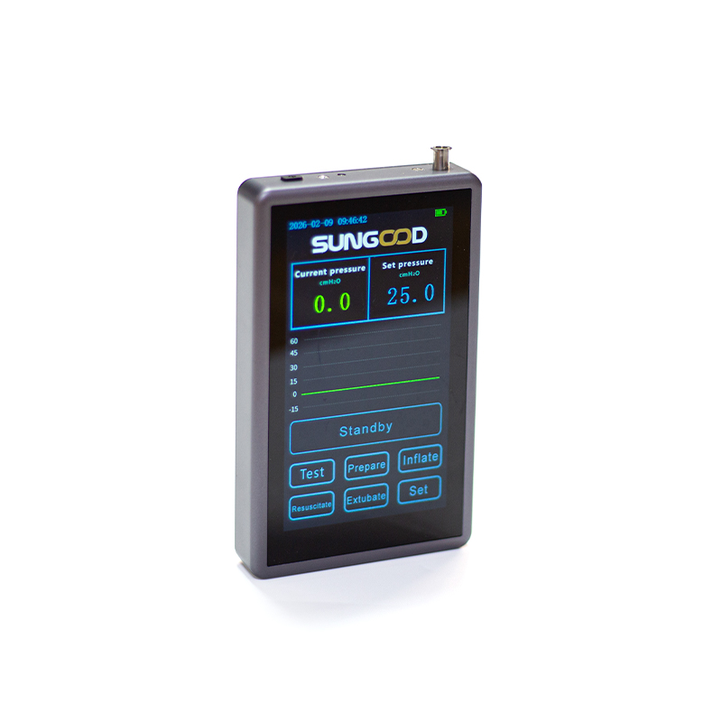
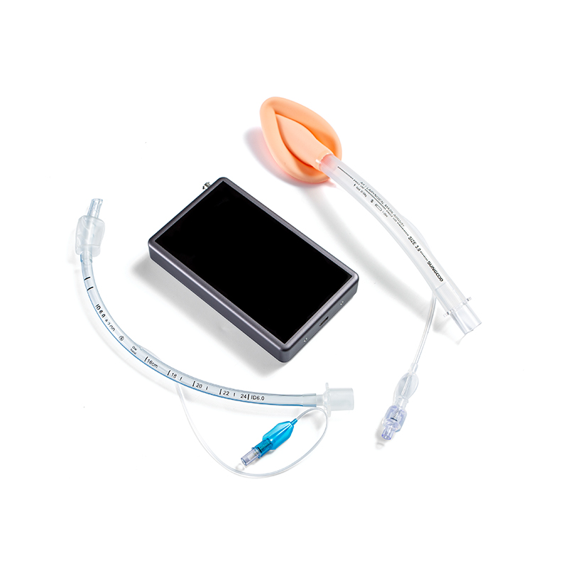 Airway Cuff Pressure Monitoring SystemContinuous cuff pressure monitoring with customizable thresholds and alarms, compatible with LMA and ETT products to support standardized airway management and reduce pressure-related risk.View Product →
A practical sequence starts with the least invasive reversible step. Open the mouth, suction visible secretions, perform a jaw thrust or head tilt–chin lift when appropriate, and remove an obvious foreign body if it can be seen and safely extracted. Listen for air movement, look for chest rise, and watch the patient’s effort. If obstruction persists, escalate to a nasopharyngeal or oropharyngeal airway, bag-mask ventilation, supraglottic airway, or tracheal intubation according to training and context.
Before inserting a device, assess three things: the likely level of obstruction, the risk of aspiration, and the expected duration of support. Supraglottic obstruction may respond to a laryngeal mask airway, but a fixed tracheal or subglottic narrowing may require a tube that passes beyond the lesion. A full stomach, active vomiting, or hematemesis raises aspiration risk and favors a cuffed tube with a seal and a suction plan. A short procedure may suit a supraglottic device, while prolonged ventilation requires a secure tracheal airway.
Cuff management is often overlooked. An overinflated cuff can injure the tracheal mucosa, while an underinflated cuff may fail to seal and allow aspiration. Measuring cuff pressure rather than judging by feel is a small step with a meaningful safety effect. For laryngeal mask airways, correct size selection and insertion technique are equally important; a device that is too small or improperly seated can leak, rotate, or obstruct. Our laryngeal mask airway sizing and safety guide covers practical insertion checks, and the respiratory and anesthesia series provides broader context for device selection.
Most common causes of upper airway obstruction are not exotic. They are tongue fallback, secretions, infection, swelling, trauma, foreign bodies, laryngospasm, tumors, and functional narrowing. The history and setting usually reveal which group is most likely, while the bedside examination shows how urgent the problem is. Stridor is a useful sign, but its absence does not rule out severe obstruction; a patient can move little air and still make little noise.
The safest approach is to name the likely mechanism early, prepare simple alternatives, and escalate before hypoxemia or complete obstruction removes the margin for error. A clear airway plan, familiar equipment, and repeated reassessment matter more than a long list of rare diagnoses.
Product Categories
Contact Us

+86-571-87687066
+86-18106505367

Workshop 1, 1st Floor(Zone B), 3rd Floor; Office Building 3rd&4th Floors, No. 550 Yinhai Street, Baiyang Sub-district, Qiantang District, Hangzhou City, Zhejiang Province, P.R.China
Ready to Begin?
Get in Touch

Now!

sungood

 English
 Español