
Home / News / Industry News / Narrowest Part of the Paediatric Airway: Glottis vs Cricoid Ring Explained
Content
A 10-month-old comes back from a 90-minute operation with a 3.5 mm tube that felt slightly snug on insertion. Two hours after extubation there is inspiratory stridor at rest, mild suprasternal recession and a rising respiratory rate. Nothing about the intubation was difficult. The problem is geometry: in a small airway, a fraction of a millimetre of mucosal swelling removes a disproportionate share of the lumen.
So where is the narrowest part of the paediatric airway? You will hear two answers, and both are defensible. Cadaver-based measurements and decades of teaching place it at the subglottic ring under the cricoid cartilage. In vivo studies using flexible bronchoscopy and MRI in living, anaesthetised children place it at the vocal cords, the same level as in adults. The first answer describes the segment with the smallest fixed diameter. The second describes the smallest diameter when the cords are open and the tissues are alive. Separate those two questions and the decisions that follow become straightforward.
The glottis can widen. The vocal cords abduct, and in most children a 4.0 mm tube passes through the same glottic opening as a 3.0 mm tube without the cords becoming the deciding factor. The subglottis cannot. The cricoid cartilage is the only complete cartilaginous ring in the airway; the thyroid cartilage and the tracheal rings are incomplete or open at the back. That single anatomical fact is why the cricoid is the textbook answer and the segment clinicians actually worry about.
It is also why the literature says "functionally narrowest portion" rather than simply "narrowest". A structure that cannot dilate, that sits in loose submucosa and that forms oedema readily is the structure that decides whether a child breathes comfortably after extubation. Subglottic stenosis, post-extubation stridor and viral croup all express themselves in the same few millimetres below the cords.
The age of the evidence matters. Older descriptions of the paediatric larynx came from cadaveric and in vitro specimens, where the cords were not abducted, muscle tone was absent and fixation had altered the soft tissues. Those specimens produced the classic picture of a funnel-shaped infant larynx with the cricoid ring at its apex. In vivo work, whether bronchoscopic or radiological, shows a subglottic region that is closer to cylindrical, with the narrowest point higher up at the glottis.
The debate is not fully closed; reviews continue to note that in vivo and in vitro findings disagree. At the bedside the disagreement matters less than it appears. Both camps agree that the subglottic region is exceptionally unforgiving, and that the margin between a comfortable airway and an obstructed one is measured in tenths of a millimetre.
Cross-sectional area scales with the square of the radius. For laminar flow, airway resistance scales with the fourth power. Take a small airway with a 3 mm radius and reduce it to 2 mm: cross-sectional area falls by roughly 55 percent, and resistance rises about fivefold. In an adult trachea of 15 to 20 mm diameter the same millimetre is a rounding error. In a neonate it is the difference between a leak and a failed extubation.
| Age group | Segment that limits you | Typical uncuffed tube ID | What to watch |
|---|---|---|---|
| Term neonate | Subglottic ring under the cricoid | 3.0 to 3.5 mm | A snug tube plus 0.5 mm of oedema can halve the lumen within hours |
| 6 to 12 months | Glottis on in vivo imaging; subglottis in practice | 3.5 to 4.0 mm | The larynx sits high and anterior, near C3 to C4 |
| Around 2 years | Glottis | 4.0 to 4.5 mm | Stridor usually appears before desaturation; act on it early |
| 2 to 8 years | Glottis | Age in years divided by 4, plus 4 mm | Half sizes matter more than the formula |
| Adolescent and adult | Glottis | 7.0 to 8.0 mm | The cricoid is no longer the limiting ring |
The narrowest segment is only one of several paediatric differences that change technique.
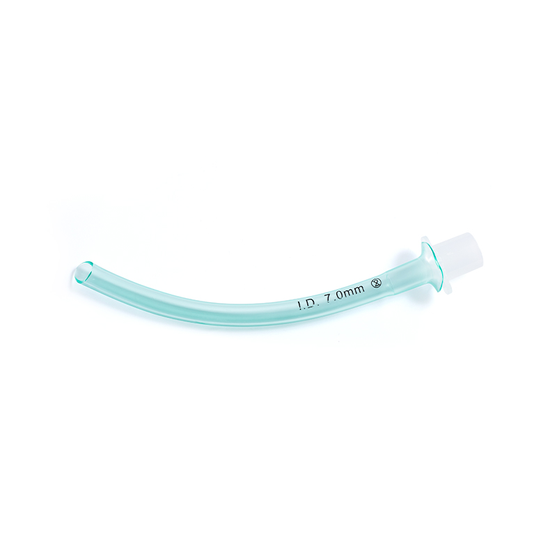
For nasal adjuncts, single-use paediatric nasopharyngeal airways supplied in clear gauge increments remove reprocessing variables from the equation entirely.
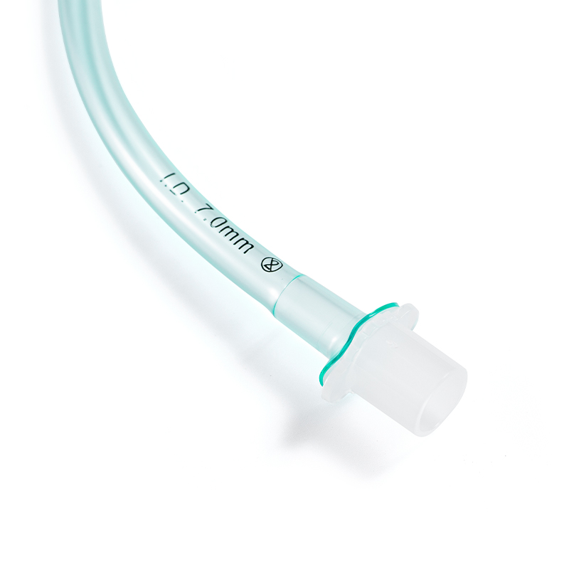 Single-Use Nasopharyngeal Airway in Gauge IncrementsSingle-use nasopharyngeal airways in paediatric sizes and clear gauge increments; the disposable design removes reprocessing variables for nasal adjunct selection.View Product →
Two rules of thumb survive scrutiny. For an uncuffed tube, internal diameter in millimetres is roughly the age in years divided by four, plus four. For a cuffed tube, deduct half a millimetre, because the cuff occupies space in the one region that cannot expand. Then confirm with a leak test: with the cuff deflated, a leak should be audible at 20 to 25 cmH2O of airway pressure. No leak means the tube is too large, and downsizing by half a millimetre is the safer response.
Cuffed or uncuffed, choose a tube for the leak rather than for a tight fit. Standard uncuffed tubes remain the default for short procedures in small children, and the subglottic fit is the reason.
 Standard Uncuffed Endotracheal Tube for Short-Term Airway SupportCuff-free standard tube for short-term anaesthesia and emergency ventilation; clear depth scale and broad neonatal-to-adult sizes support leak-based sizing.View Product →
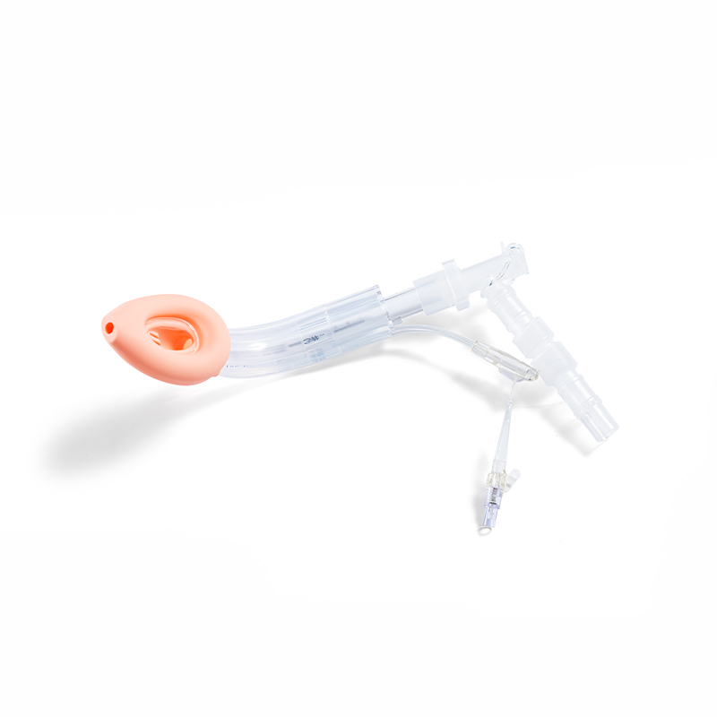
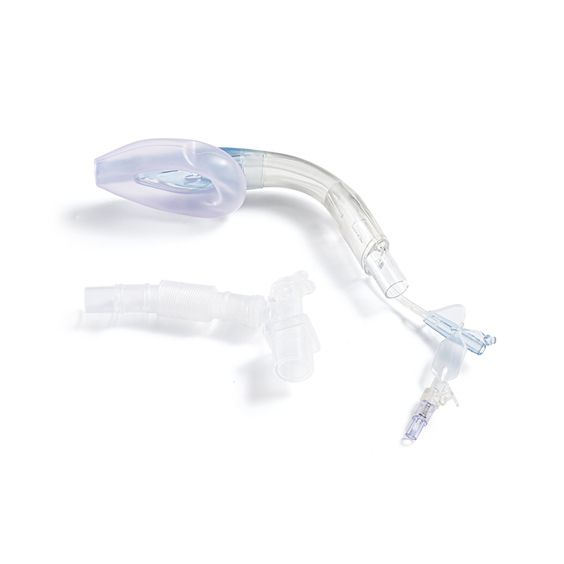
Where the subglottis is a known concern, a supraglottic airway sidesteps it, because the device never passes the cords. The trade-off is seal pressure and aspiration protection, which is why variants with gastric access channels exist. Sizing follows weight rather than age, in small increments: approximately size 1 below 5 kg, 1.5 from 5 to 10 kg, 2 from 10 to 20 kg, 2.5 from 20 to 30 kg and 3 from 30 to 50 kg. Insertion and sizing have their own learning curve, so the practical guide to laryngeal mask insertion and sizing is worth reading before the first paediatric case in a new unit.
Paediatric airway ranges are judged by their gaps rather than their headliners. A range that jumps from 3.0 mm to 4.0 mm is not a paediatric range. Half sizes, clear depth markings, a Murphy eye and a low-profile cuff are what prevent the scenario described at the start of this article.
For supraglottic devices, weight-based sizes and a documented maximum seal pressure matter more than any claim about softness. A silicone mask that covers the weight bands your unit actually uses is worth more than a broad range that skips one of them.
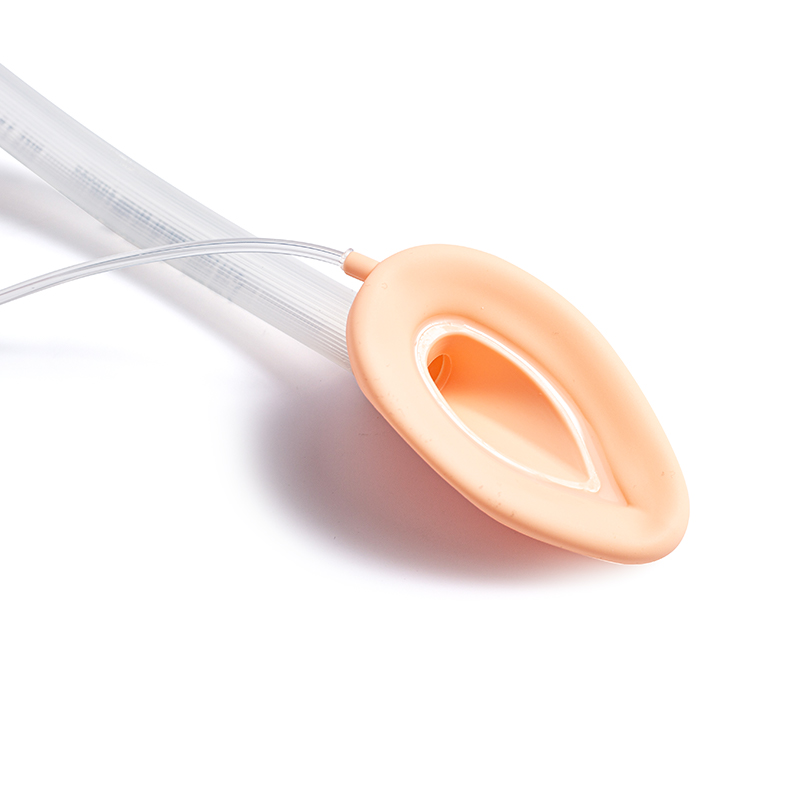 Standard Silicone Laryngeal Mask AirwaySilicone supraglottic airway with weight-based sizes from neonates to adults; covers common paediatric and adult weight bands for anaesthesia, resuscitation and ICU ventilation.View Product →
Materials deserve a second look. PVC holds its shape at body temperature; silicone is softer and more forgiving of an imperfect angle but behaves differently as it warms. For nasal and gastric devices, a fully radiopaque stripe makes post-insertion checks faster and less ambiguous. Claims about manufacturing conditions, whether a 100,000-class GMP cleanroom, CE marking or ISO 13485 certification, should be verifiable in documentation rather than accepted from a product page, because in paediatric airway devices a problem tends to surface as a batch-level failure rather than a design-level one.
If you are comparing suppliers, ask for samples in the half sizes you actually use before committing to a volume order. Contact us to request samples, dimensional data or a quotation for the sizes your unit stocks.
Decide which question you need answered. If you are asking which segment has the smallest diameter in a living child, the answer is the glottis at the level of the vocal cords. If you are asking which segment determines whether that child breathes comfortably six hours after extubation, the answer is the subglottic ring beneath the cricoid cartilage, the one part of the paediatric airway that will not give. Size to the subglottis, confirm with a leak test at 20 to 25 cmH2O, keep half sizes on the shelf, and treat stridor after extubation as a problem to solve rather than a noise to observe.
Product Categories
Contact Us

+86-571-87687066
+86-18106505367

Workshop 1, 1st Floor(Zone B), 3rd Floor; Office Building 3rd&4th Floors, No. 550 Yinhai Street, Baiyang Sub-district, Qiantang District, Hangzhou City, Zhejiang Province, P.R.China
Ready to Begin?
Get in Touch

Now!

sungood

 English
 Español