
Home / News / Industry News / Vacuum Blood Collection Tube Guide: Types, Order of Draw & Errors
Content
The accuracy of laboratory diagnostics hinges on the pre-analytical phase, where the vacuum blood collection tube plays a pivotal role. These tubes are engineered with specific additives and precise vacuum levels to ensure the correct blood-to-additive ratio, which is critical for valid test results. Improper selection or handling of these tubes accounts for nearly 70% of all pre-analytical errors in clinical laboratories. This guide details the functional differences between tube types, the mandatory order of draw, and practical techniques to minimize sample rejection due to hemolysis or clotting.
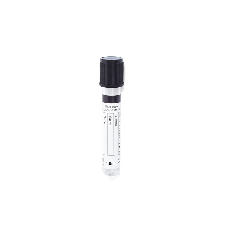
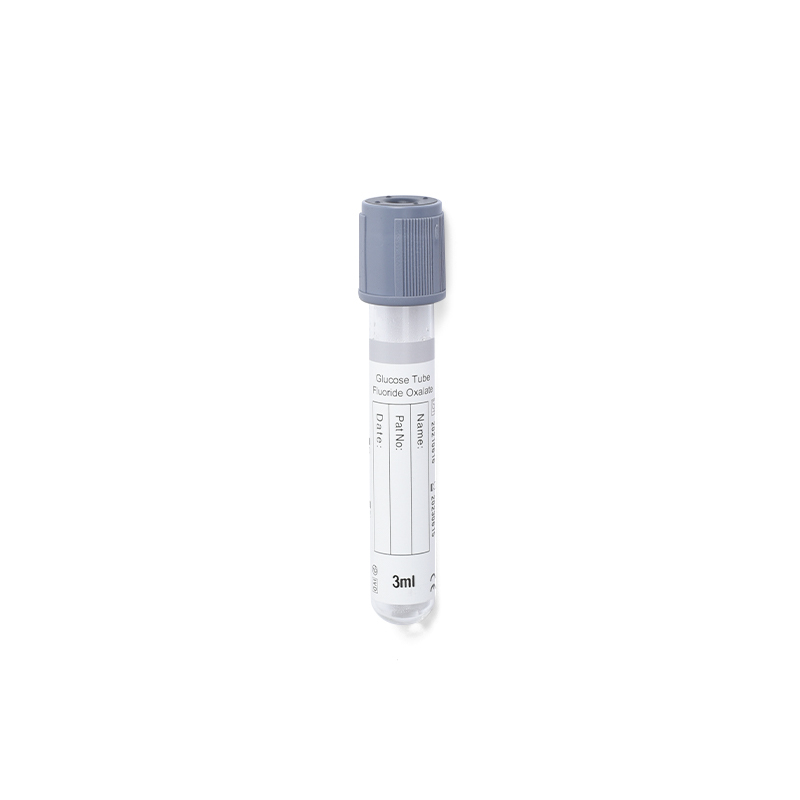
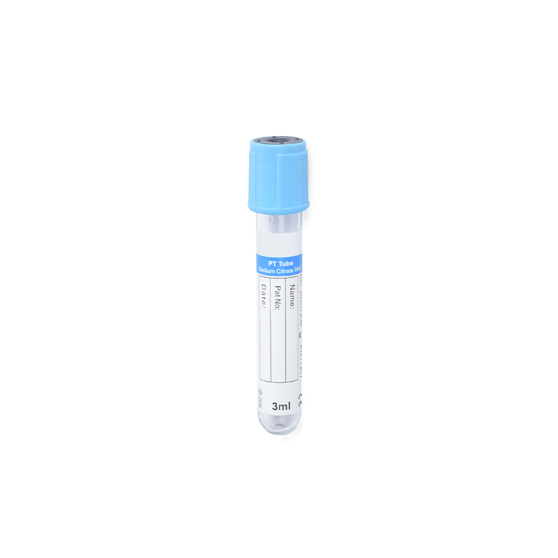
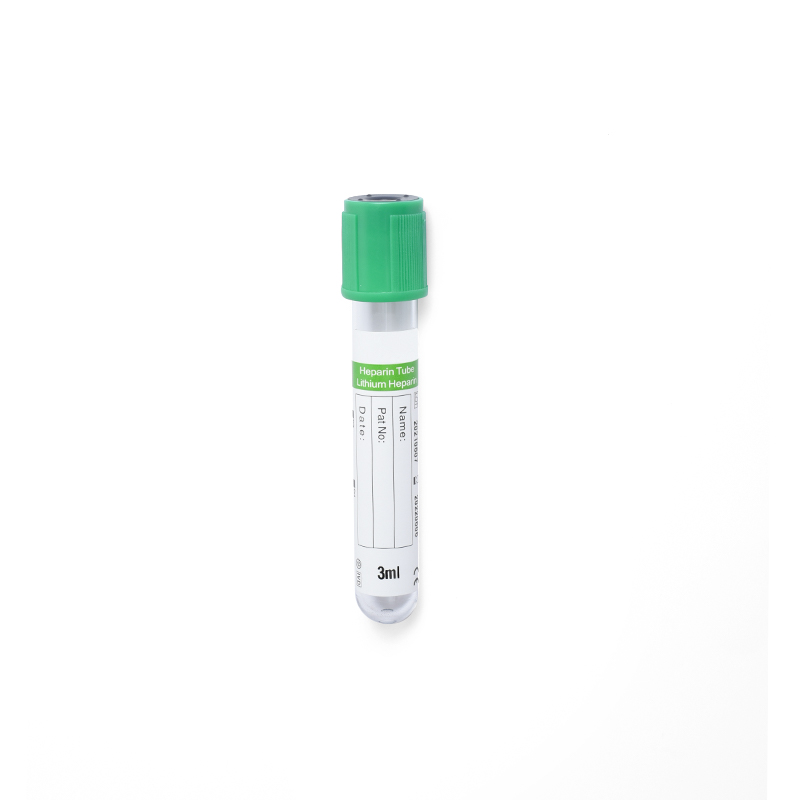
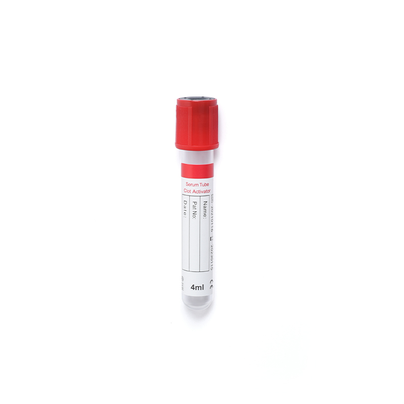
Each vacuum blood collection tube color corresponds to a specific chemical additive that preserves the blood sample for particular tests. Using the wrong tube can lead to catastrophic analytical interference, such as potassium leakage from cells or calcium chelation.
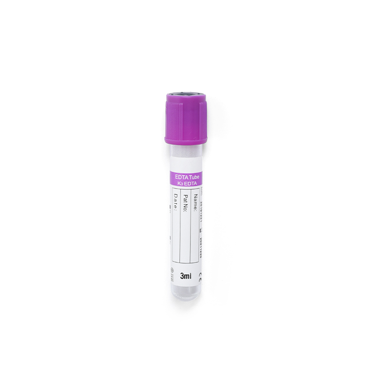
Understanding the mechanism of each additive helps in troubleshooting sample issues. For instance, EDTA works by chelating calcium to prevent clotting, making it ideal for hematology but useless for coagulation studies.
| Tube Color | Additive | Primary Use | Key Inversion Count |
|---|---|---|---|
| Light Blue | Sodium Citrate | Coagulation (PT/INR, PTT) | 3-4 times |
| Red/Gold | Clot Activator/SST | Chemistry, Serology, Immunology | 5 times |
| Green | Heparin (Li/Na) | Stat Chemistry, Plasma | 8-10 times |
| Lavender | EDTA (K2/K3) | Hematology (CBC), Blood Bank | 8-10 times |
| Gray | Sodium Fluoride | Glucose Tolerance, Alcohol | 8-10 times |
The order in which vacuum blood collection tubes are filled is not arbitrary; it is designed to prevent cross-contamination of additives between tubes. Carryover of even microscopic amounts of additive can skew results significantly. For example, EDTA carryover into a chemistry tube will cause falsely low calcium and high potassium readings.
Deviation from this order is a leading cause of sample rejection in high-volume laboratories. Always follow the specific protocol provided by your laboratory, as some institutional guidelines may vary slightly based on testing platforms.
Hemolysis, the rupture of red blood cells, releases intracellular contents into the serum or plasma, interfering with many assays. It is the most common reason for specimen rejection. Proper technique with the vacuum blood collection tube is essential to prevent this.
Once the blood is collected, the stability of the analytes depends on proper storage and transport conditions. Delays in processing can alter glucose levels, gas tensions, and cell morphology.
Time Limits: Serum or plasma should generally be separated from cells within 2 hours of collection. If immediate separation is not possible, store whole blood at room temperature for coagulation tests and refrigerated (2-8°C) for most chemistry tests, unless specified otherwise.
Light-sensitive analytes, such as bilirubin and vitamin B12, require tubes to be wrapped in aluminum foil or placed in amber-colored vacuum blood collection tubes to prevent photodegradation. Always verify transport requirements with the receiving laboratory to ensure sample integrity upon arrival.
Product Categories
Contact Us

+86-571-87687066
+86-18106505367

Workshop 1, 1st Floor(Zone B), 3rd Floor; Office Building 3rd&4th Floors, No. 550 Yinhai Street, Baiyang Sub-district, Qiantang District, Hangzhou City, Zhejiang Province, P.R.China
Ready to Begin?
Get in Touch

Now!
sungood
 English
 Español