
Home / News / Industry News / Endotracheal Tube: Selection, Safety & Clinical Best Practices
Content
The endotracheal tube (ETT) is the definitive airway device in critical care and anesthesia, serving as the primary conduit for ventilation and protection against aspiration. Effective management of an ETT is not merely about insertion; it requires rigorous adherence to safety protocols to prevent life-threatening complications such as ventilator-associated pneumonia (VAP) and laryngeal trauma. The most critical operational parameter is maintaining cuff pressure between 20 and 30 cm H2O; pressures below this range increase aspiration risk, while pressures above it can cause tracheal mucosal ischemia and permanent damage.
This article provides a comprehensive guide to selecting the appropriate ETT, managing cuff dynamics, and implementing evidence-based practices to minimize complications. By understanding these technical and clinical nuances, healthcare providers can ensure optimal patient outcomes during mechanical ventilation.
Choosing the correct endotracheal tube is the first step in securing a safe airway. The decision depends on patient anatomy, gender, and the specific clinical context, such as surgical requirements or anticipated difficult intubation.
For adult patients, the internal diameter (ID) is typically selected based on gender and height. The standard ID for an average adult female is 7.0–7.5 mm, while for an average adult male, it is 8.0–8.5 mm. Using a tube that is too small increases airway resistance and makes suctioning difficult, whereas a tube that is too large can cause vocal cord injury and post-extubation stridor. In pediatric patients, sizing is often calculated using the formula: ID = (Age/4) + 4 for cuffed tubes.
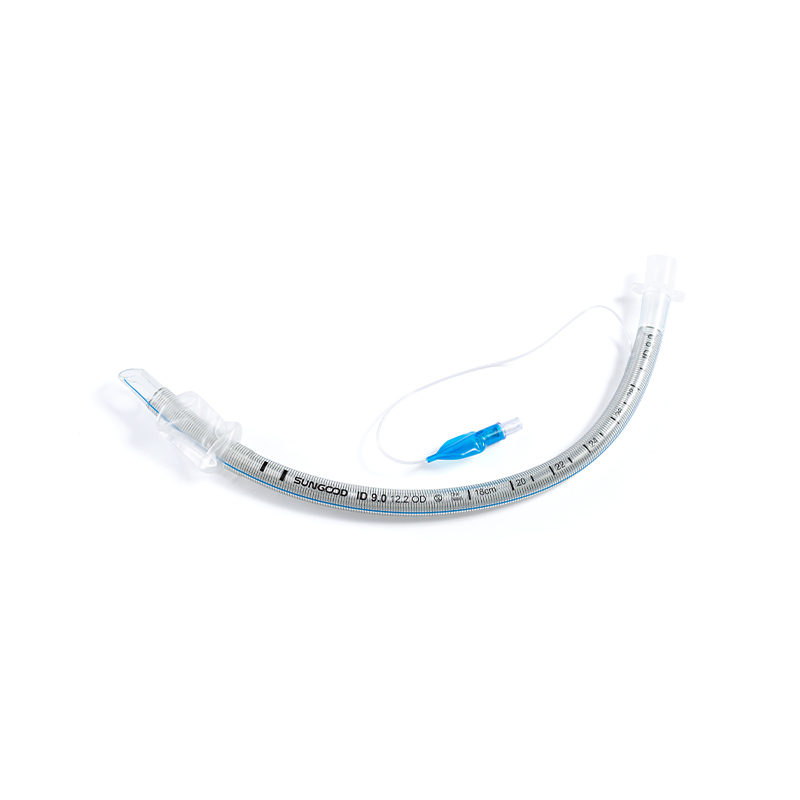
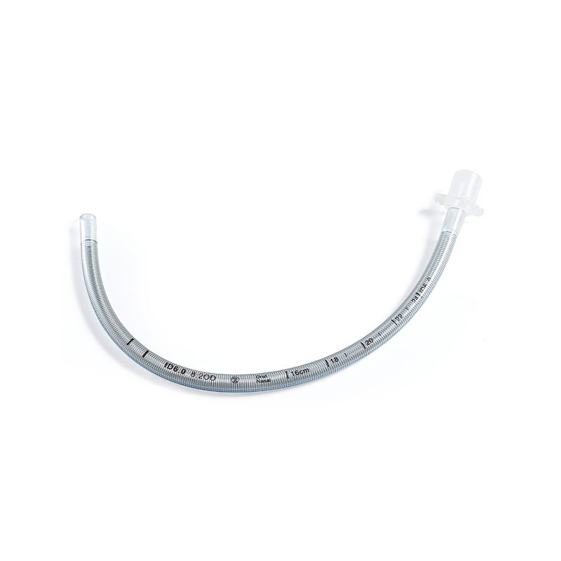
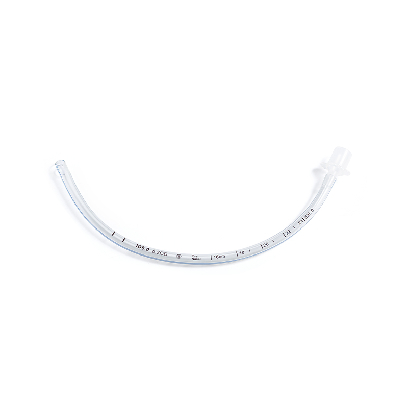
Standard PVC tubes are common, but specialized designs offer distinct advantages. Microcuff tubes feature a thinner polyurethane cuff that seals more effectively at lower pressures, reducing the risk of microaspiration. Reinforced (armored) tubes resist kinking and are essential for head and neck surgeries where the tube may be bent. Selecting the right type ensures both patency and safety throughout the procedure.
| Patient Group | Typical ID (mm) | Insertion Depth (cm) | Key Consideration |
|---|---|---|---|
| Adult Female | 7.0 – 7.5 | 21 – 23 | Smaller larynx structure |
| Adult Male | 8.0 – 8.5 | 22 – 24 | Larger tracheal diameter |
| Child (1-8 yrs) | (Age/4) + 4 | Depth = 3 x ID | Cricoid ring is narrowest point |
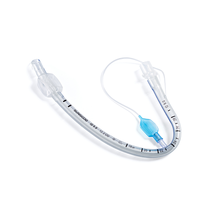
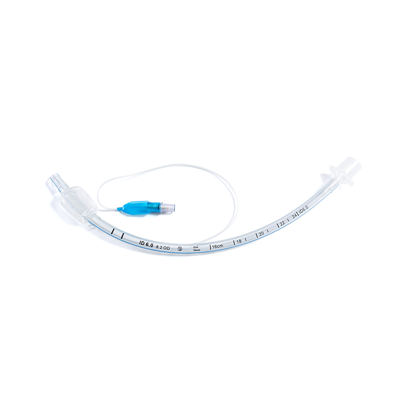
The inflatable cuff on an endotracheal tube serves two vital functions: it seals the airway to allow for positive pressure ventilation and prevents oral secretions from entering the lungs. Improper cuff management is a leading cause of iatrogenic tracheal injury.
Tracheal mucosal capillary perfusion pressure is approximately 20–30 cm H2O. When cuff pressure exceeds this threshold, blood flow to the tracheal wall is obstructed, leading to ischemia, ulceration, and potentially tracheomalacia or stenosis. Studies indicate that up to 50% of intubated patients have cuff pressures outside the safe range if not monitored regularly. Therefore, continuous or intermittent monitoring using a manometer is mandatory.
Conversely, low cuff pressure allows contaminated oropharyngeal secretions to leak past the cuff into the lower airways, a phenomenon known as microaspiration. This is a primary pathway for the development of ventilator-associated pneumonia (VAP). Maintaining the minimum occluding volume or a pressure of at least 20 cm H2O is essential to create an effective seal without compromising mucosal integrity.
Prolonged intubation carries risks beyond cuff-related injuries. Implementing bundled care strategies significantly reduces the incidence of VAP, accidental extubation, and sinusitis.
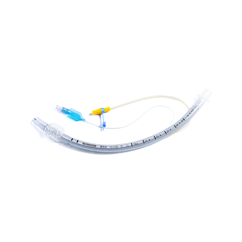
Secretions pool above the ETT cuff, creating a reservoir of bacteria. Endotracheal tubes with dorsal subglottic suction ports allow for the continuous or intermittent removal of these secretions. Clinical trials have shown that subglottic suctioning can reduce the incidence of VAP by up to 50% in patients expected to be intubated for more than 72 hours.
Rigorous oral hygiene with chlorhexidine gluconate reduces bacterial load in the oropharynx. Additionally, securement devices should be used to prevent tube movement, which can cause laryngeal abrasion. Rotating the tube position slightly (if tolerated and protocol allows) and alternating oral care sides can prevent pressure ulcers on the lips and commissures. Daily sedation vacations and readiness-to-wean assessments help minimize the duration of intubation, thereby reducing cumulative risk.
Product Categories
Contact Us

+86-571-87687066
+86-18106505367

Workshop 1, 1st Floor(Zone B), 3rd Floor; Office Building 3rd&4th Floors, No. 550 Yinhai Street, Baiyang Sub-district, Qiantang District, Hangzhou City, Zhejiang Province, P.R.China
Ready to Begin?
Get in Touch

Now!

sungood

 English
 Español