
Home / News / Industry News / Endotracheal Tube Guide: Types, Sizing & Clinical Best Practices
Content
An endotracheal tube is a critical medical device used to establish and maintain a patent airway, ensuring effective ventilation and oxygenation in patients who cannot breathe adequately on their own. Proper selection, insertion, and management of the tube are essential to prevent complications such as hypoxia, aspiration, and tracheal damage.
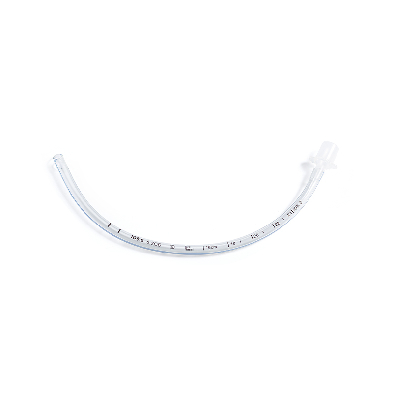
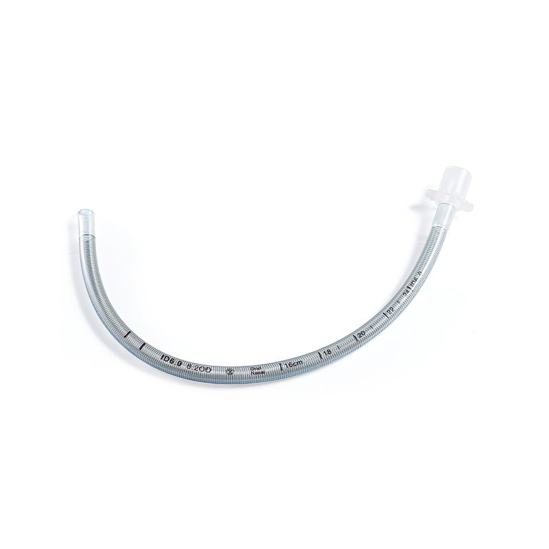
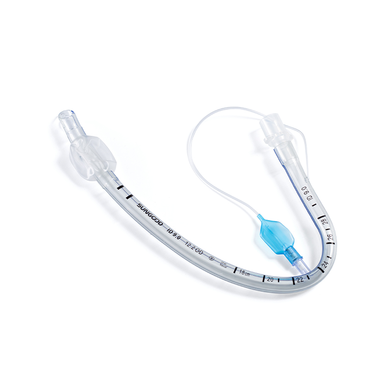
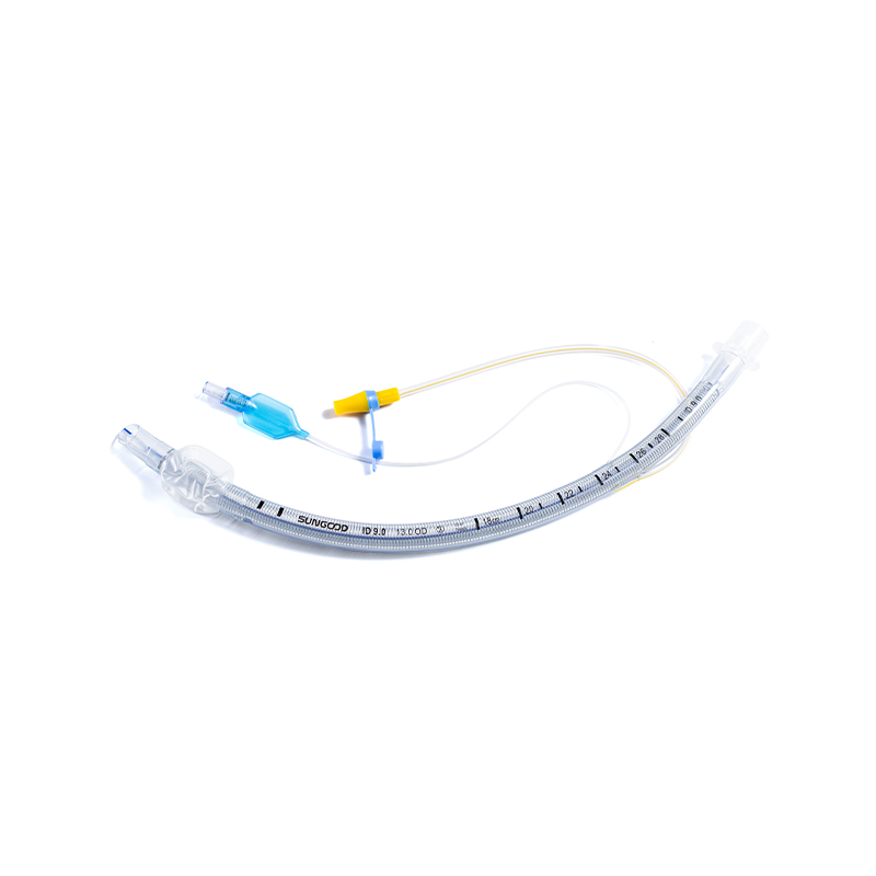
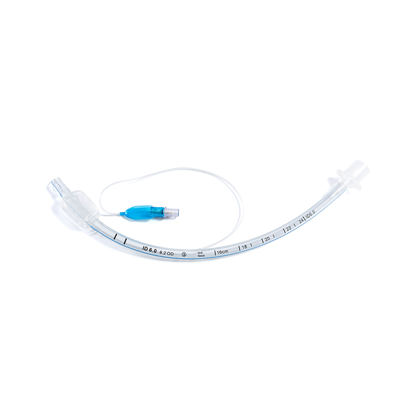
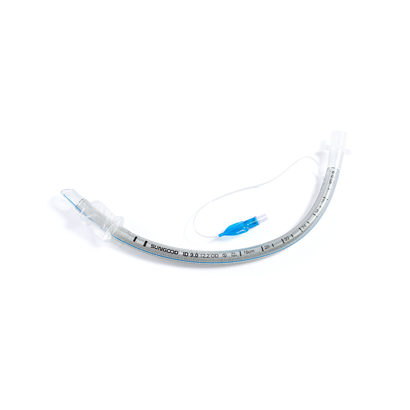
Understanding the structural components of an endotracheal tube (ETT) is fundamental for safe clinical application. Each part serves a specific function to facilitate intubation and secure the airway.
The Murphy eye, a hole on the side of the tube tip, provides an alternative airflow path if the main lumen becomes obstructed by secretions or the tracheal wall, enhancing patient safety during prolonged intubation.
Choosing the appropriate internal diameter (ID) is crucial for minimizing airway resistance while preventing tracheal injury. Incorrect sizing can lead to significant complications, including post-extubation stridor or inadequate ventilation.
| Patient Group | Internal Diameter (mm) | Insertion Depth (cm) |
|---|---|---|
| Adult Female | 7.0 – 8.0 | 21 – 23 |
| Adult Male | 8.0 – 9.0 | 22 – 24 |
| Child (1-10 yrs) | (Age/4) + 4 | Depth = ID x 3 |
| Infant (<1 yr) | 3.0 – 4.5 | 7 – 10 |
For adult males, an 8.0 mm ID tube is generally preferred as it balances low airflow resistance with acceptable trauma risk. In contrast, smaller tubes (≤7.0 mm) significantly increase work of breathing due to higher resistance, which can complicate weaning from mechanical ventilation.
The presence of a cuff distinguishes most adult endotracheal tubes. The decision to use a cuffed or uncuffed tube depends on the clinical context and patient age.
Cuffed tubes provide a seal against the tracheal wall, allowing for positive pressure ventilation without significant air leak. This seal is critical for protecting the lungs from aspiration of blood, vomit, or secretions, making cuffed tubes the standard for emergency and surgical airways in adults.
Historically, uncuffed tubes were used in children under 8 years old due to the narrowest part of the pediatric airway being the cricoid ring. However, modern micro-cuff tubes are increasingly used in pediatrics to allow for better ventilation control and reduced need for tube exchange, provided cuff pressures are strictly monitored below 20-25 cm H2O.
Intubation and prolonged use of an endotracheal tube carry risks. Awareness of potential complications enables proactive management and improved patient outcomes.
Prolonged intubation can lead to laryngeal edema, vocal cord paralysis, or tracheal stenosis. Maintaining cuff pressure between 20 and 30 cm H2O is vital to prevent tracheal mucosal ischemia, which can result in necrosis and subsequent stenosis.
Proper care of the endotracheal tube reduces the risk of accidental extubation and ventilator-associated pneumonia (VAP).
Adhering to these evidence-based practices ensures that the endotracheal tube serves its life-saving purpose while minimizing harm to the patient.
Product Categories
Contact Us

+86-571-87687066
+86-18106505367

Workshop 1, 1st Floor(Zone B), 3rd Floor; Office Building 3rd&amp;4th Floors, No. 550 Yinhai Street, Baiyang Sub-district, Qiantang District, Hangzhou City, Zhejiang Province, P.R.China
Ready to Begin?
Get in Touch

Now!
sungood
 English
 Español